Posts

Patricia J. Johnson

UCLA microbiologist Patricia J. Johnson elected to National Academy of Sciences
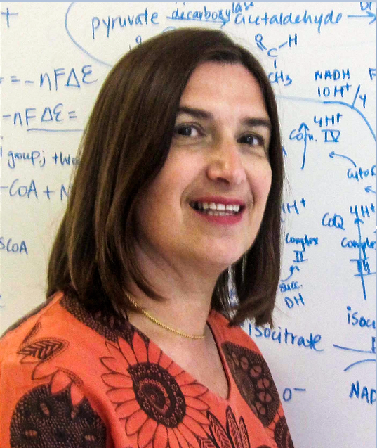
Patricia J. Johnson

Patricia J. Johnson

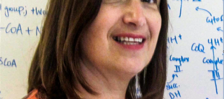
Patricia J. Johnson, UCLA professor of microbiology, immunology and molecular genetics, was elected to the National Academy of Sciences in recognition of her “distinguished and continuing achievements in original research.”

Membership in the academy is one of the highest honors that a U.S. scientist can receive. Its members have included Albert Einstein, Thomas Edison, Orville Wright and Alexander Graham Bell. The academy today announced the election of 100 new members and 25 foreign associates.

“I am very honored to be include among the ranks of such distinguished scientists,” said Johnson, who has appointments in the David Geffen School of Medicine at UCLA and the division of life sciences in the UCLA College.

Research in Johnson’s laboratory focuses on the molecular and cellular biology of a single cellular parasite called Trichomonas vaginalis. This microbe is responsible for the most prevalent, non-viral, sexually transmitted infection worldwide and is the most common parasite found in the U.S. population. An estimated 275 million people worldwide have the parasite, including approximately 3.7 million in the United States. In 2014, the Centers for Disease Control and Prevention identified trichomoniasis, the infection caused by T. vaginalis, as one of the “neglected parasitic infections in the United States.”

Johnson said that beyond its medical importance, T. vaginalis is a fascinating organism for conducting research on the evolution of biological processes present in all eukaryotes, from microbes to humans. The parasite’s atypical properties offer possible chemotherapeutic targets and vaccine candidates, she said.

Her laboratory focuses on several aspects of trichomonad biology, including its evolution, regulation of gene expression, drug resistance, genomics and biological processes vital for human infection.

“Our interdisciplinary research program merges several specialties, including structural and cell biology, biochemistry, genomics, proteomics, bioinformatics, evolution and medical sciences,” she said. “In recent years, we have narrowed our focus to defining and explaining critical pathogenic mechanisms that allow T. vaginalis to establish and maintain an infection. These studies include identifying critical parasite cell surface molecules and secreted vesicles, as well as defining human immune responses to parasitic infection. We have also investigated a possible link between infection with T. vaginalis and prostate cancer.”

The National Academy of Sciences was established in 1863 by a congressional act of incorporation signed by Abraham Lincoln that calls on the academy to act as an official adviser to the federal government, upon request, in any matter of science or technology. The academy is a private organization of scientists and engineers dedicated to the furtherance of science and its use for the general welfare.

Photo of UCLA professor Kent Hill and graduate student Stephanie DeMarco

Scientists identify a key gene in the transmission of deadly African sleeping sickness

Photo of UCLA professor Kent Hill and graduate student Stephanie DeMarco

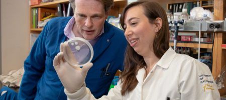
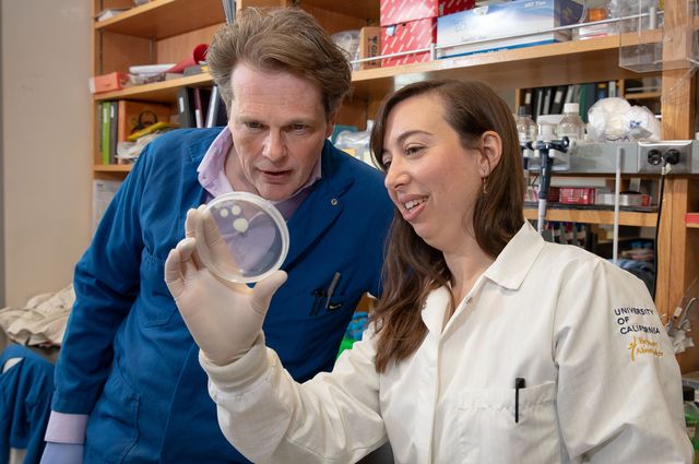
Research by UCLA professor Kent Hill and graduate student Stephanie DeMarco, as well as colleagues at the University of Bern, could lead to new approaches to treat African sleeping sickness.


Life scientists from UCLA and the University of Bern have identified a key gene in the transmission of African sleeping sickness — a severe disease transmitted by the bite of infected tsetse flies, which are common in sub-Saharan Africa.

The disease is fatal if untreated, as the parasite responsible moves from the bloodstream to the central nervous system. Tens of millions of people in 36 African countries are at risk. There is no vaccine, and conventional drug treatments, which include an arsenic derivative, are antiquated, not very effective and have severe side effects.

The research, published in the journal Nature Communications, could lead to new approaches to treat the disease. It also provides scientists with the first detailed understanding of how the parasite moves through the fly and what genes enable it to do so.

The tiny, single-celled parasite that causes African sleeping sickness in humans, and debilitating diseases in other mammals, is called Trypanosoma brucei, or T. brucei. To become infectious, the parasite must travel through tissues of the fly, from the midgut to the salivary gland — and then into the human or other animal, through a bite.

In the study, Stephanie DeMarco, a UCLA graduate student in molecular biology, and Sebastian Shaw, a graduate student at Switzerland’s University of Bern, worked with two sets of the T. brucei parasite. In one set, they made a mutation in one of the parasite’s genes, called phosphodiesterase-B1, or PDEB1.

Then, they infected 2,000 tsetse flies with some 20,000 parasites each — half of the flies received blood containing normal T. brucei parasites and the other half received blood with the mutated versions.

When tsetse flies drink infected blood, the parasites from the blood typically travel to the midgut and then into a tissue closer to the head, called the proventriculus, before moving on to the salivary glands.

But the researchers saw a striking difference in the proventriculus between the two sets of flies. Among the flies that received the normal parasites, those that had parasites in the gut also had parasites in the proventriculus; but among the 1,000 flies that received mutant T. brucei, only a single one that had parasites in the gut also had a parasite in the proventriculus.

“The normal parasites were able to get to the proventriculus just fine, but for the mutants, we saw only one lonely parasite swimming around,” DeMarco said. “That told us that phosphodiesterase-B1 is really important for the parasites to move from the fly midgut to the proventriculus.”

Shaw said, “When we saw the huge difference between the mutants and normal parasites, at first we couldn’t believe it.”

Kent Hill, a UCLA professor of microbiology, immunology and molecular genetics, and one of the study’s senior authors, said the findings also suggested that there must be a barrier preventing the mutants from getting from the midgut to the proventriculus.

To learn where that barrier is, the scientists made fluorescent parasites and fed the flies a fluorescent dye that stained different tissues in the fly different colors, enabling the researchers to track the parasites.

To go from the midgut to the proventriculus, the parasites have to cross the peritrophic matrix, a sheet-like structure produced by the proventriculus that protects the midgut.

“We found the normal parasites could get through the peritrophic matrix just fine, but the mutants were mostly stuck on one side of it,” DeMarco said.

That finding indicated that the peritrophic matrix was the barrier the scientists were looking for.

The research identifies for the first time the genes that enable the parasites to sense where they are and allow them to survive their journey in fly tissues; those mechanisms had not been understood well until now.

“We think the way the parasites perceive where they are may be similar in the tsetse flies and in mammals — including humans — as they go through barriers and tissues,” said co-senior author Isabel Roditi, a University of Bern professor. “If so, there could potentially be a new drug that might disrupt their ability to do that.”

The researchers also uncovered another clue to African sleeping sickness: In parasites with mutated PDEB1, there was a dramatic increase in the number of cyclic AMP molecules, signaling molecules that play an important role in the disease.

Normal parasites are social and coordinate their behavior, DeMarco said. But the research revealed that without PDEB1, the parasites have too much cyclic AMP in their cells and can’t communicate with one another.

“When Sebastian and Stephanie got rid of PDEB1, the parasites got flooded with cyclic AMP,” Hill said. “Then, when the signal came in telling the parasites, ‘You’re in the stomach and you need to move,’ they couldn’t hear the sound. That’s what we think the problem is for the mutant parasites.”

Hill said the new insights from the UCLA–Bern study could apply to other disease-causing parasites as well. For example, T. brucei parasites are related to parasites found in the U.S. and elsewhere that cause Chagas disease, in which parasites invade heart tissue, leading to inflammation and enlarged heart tissue, and in some cases, heart failure.

Hill’s research is funded by the National Institutes of Health’s National Institute of Allergy and Infectious Diseases and the National Institute of General Medical Sciences. Roditi’s research is funded by the Swiss National Science Foundation and the Howard Hughes Medical Institute.

Photo of Mark Pollock and Simone George

UCLA scientist gives couple hope while searching for a cure for paralysis

Photo of Mark Pollock and Simone George

Mark Pollock and Simone George

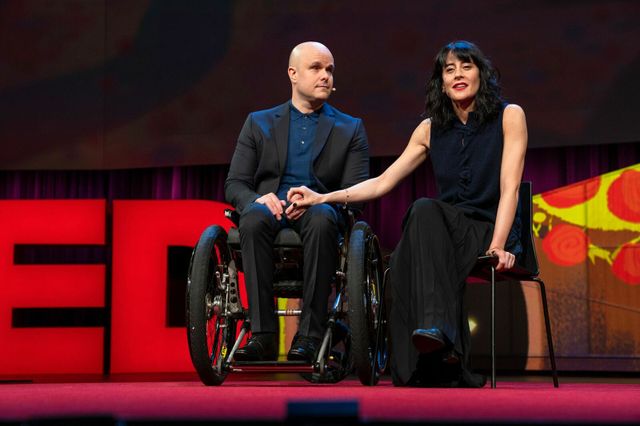
If you think listening to a paralyzed, blind man discuss his life does not sound uplifting, meet Mark Pollock. Social psychologist Amy Cuddy, whose TED talk about body language has nearly 15 million views, describes the new talk by Pollock and his partner, human rights lawyer, Simone George, as “the most powerful, moving talk I have ever seen at TED.”

Pollock lost his vision at 22 and became paralyzed after an accident at 39 that left him, in his words, “on the very edge of survival.” You certainly wouldn’t know that seeing him now in this talk, titled “A love letter to realism in a time of grief,” and the fulfilling life he is living.

In the talk, he and George discuss how during the 16 months spent in hospitals, doctors discouraged them from hoping for a cure for paralysis.

“Cancelling hope ran contrary to everything that we believed in,” says Pollock, who in 2009 became the first blind person to race to the South Pole. “Yes, up to this point in history, it had proven to be impossible to find a cure for paralysis, but history is filled with accounts of the impossible made possible through human endeavor — the kind of human endeavor that took explorers to the South Pole at the start of the last century, and the kind of human endeavor that will take adventurers to Mars in the early part of this century. We started asking why can’t that same kind of human endeavor cure paralysis in our lifetime.”

A robotic exoskeletal device allowed Pollock to stand and walk more than 1 million steps, although George explains that the robotic device was doing all the work.

Then George says in the talk, they met a “true visionary” at UCLA: Dr. Reggie Edgerton, “the most beautiful man” whose “life work had resulted in a scientific breakthrough.” Edgerton is a UCLA distinguished professor of integrative biology and physiology, neurobiology and neurosurgery.

“Using electrical stimulation of the spinal cord, a number of subjects had been able to stand and regain some movement and feeling — and most importantly, to regain some of the body’s internal functions that are designed to keep us alive and to make that life a pleasure,” George says. “Electrical stimulation of the spinal cord, we think, is the first meaningful therapy ever for paralyzed people.”

Pollock is the first person with chronic, complete paralysis to regain enough voluntary control to actively work with a robotic device and take steps. Applying electrical stimulation of his spinal cord as he walked in his robotic exoskeletal device was like watching Iron Man, George says.

“Simone, my robot and I moved into the lab at UCLA for three months,” Pollock says. “Every day, Reggie and his team put electrodes onto the skin on my lower back and pushed electricity into my spinal cord to excite my nervous system as I walked in my exo, and for the first time since I was paralyzed, I could feel my legs underneath me.”

Because of the stimulation, he was able to voluntarily move his paralyzed legs. “As I did more, the robot intelligently did less,” he says. “My heart rate got to a normal running training zone of 140 to 160 beats per minute, and my muscles, which had almost entirely disappeared, started to come back.”

Lying on his back in Edgerton’s laboratory more than three-and-a-half years after he became paralyzed, Pollock was able, with the electrical stimulator turned on, to pull his knee to his chest.

“Science is love,” George says. The two of them have discarded the advice from the earlier doctors to avoid hoping for a cure.

A theme of their TED talk is how to resolve the tension between acceptance and hope.

Sixty million people worldwide are paralyzed. Pollock’s life inspires hope to them and many others.

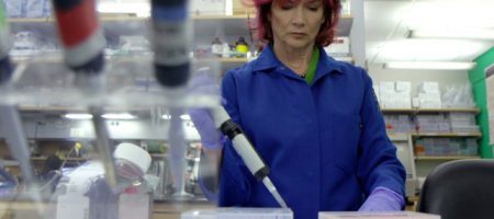
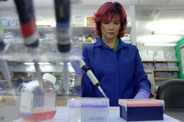
Rachelle Crosbie-Watson in her lab at UCLA.

Curing a deadly childhood disease, sharing her love of science, and a sleek ’68 Corvette drive this biochemist

Rachelle Crosbie-Watson in her lab at UCLA.

Rachelle Crosbie-Watson in her lab at UCLA.


Spend a brief amount of time with biochemist Rachelle Crosbie-Watson and you’ll quickly realize that “drive” is one of her favorite words.

With equal enthusiasm, she’ll describe studying “the small molecules that drive life,” and her 1968 convertible Corvette being “a blast to drive.”

The symmetry is hard to miss: Crosbie-Watson drives a classic muscle car to UCLA, where she studies the biochemical reactions that drive muscle cell functions. Her lab is hotly pursuing new drugs that one day may halt the progression of a deadly childhood muscle-wasting disease, allowing kids with the disorder to lead normal lives.

The popular digital network, Mashable, recently profiled Crosbie-Watson for its “How She Works” series, which shadows a day in the life of women professionals working in fields related to science, technology, engineering and math, or STEM.

With her fiery pink hair, charismatic personality and affinity for high-speed cars, Crosbie-Watson doesn’t resemble most people’s vision of a biochemist. But her talent for crafting fresh approaches to solving thorny scientific puzzles is exactly what makes her such an ingenious scientist.

“What I love most about my job is the opportunity to be creative,” Crosbie-Watson said. “To solve the biggest problems in the world, we need individuals with different viewpoints to chime in. Working with people who are learning science for the first time — coupled with the thrill of discovery — makes for a really exciting recipe.”

Crosbie-Watson wears a lot of hats. Starting July 1, she will chair the integrative biology and physiology department in the UCLA College. She is also a professor of neurology in the David Geffen School of Medicine at UCLA, and the education liaison for the Center for Duchenne Muscular Dystrophy at UCLA.

In a sunny space in the Terasaki Life Sciences Building, Crosbie-Watson oversees a window-lined laboratory staffed by young researchers. Reflecting her appeal as a mentor and role model, 14 of the 17 are female.

Her team is intent on finding a cure for Duchenne muscular dystrophy, a deadly genetic disease that slowly weakens every muscle of the body. Striking 1 in 5,000 boys, the disorder typically reveals itself in frequent falls near age 4, reliance on a wheelchair by age 12, and teenage loss of the ability to move the upper arms. Young men with Duchenne frequently die in their 20s, when their heart and lung muscles stop pumping, leading to organ failure.

“Duchenne is a horrible disease that steals young boys’ childhoods and takes young men in the primes of their lives,” Crosbie-Watson said.

The disorder is caused by a genetic error that blocks the production of dystrophin, a protein that normally protects the membrane around muscle cells as they contract and relax. Left susceptible to damage from daily wear and tear, the unprotected cells eventually begin leaking their contents into the surrounding tissue, progressively weakening the muscle until it stops working.

Her lab’s earlier studies in mice gave Crosbie-Watson an insight into how to halt that process.

“We found that boosting levels of a molecule called sarcospan restored the membrane’s ability to protect muscle cells,” she said. “Sarcospan strengthens the muscle’s capacity to withstand the forces of daily use, diminishing the harm caused by Duchenne.”

Led by graduate student Cynthia Shu, the lab began scanning thousands of potential drugs to identify ones able to elevate cellular levels of sarcospan. Three years and 200,000 candidates later, the team has identified a handful of promising contenders for preclinical testing.

Crosbie-Watson applies the same imaginative approach she follows in research to her teaching. To educate the next generation of scientists about Duchenne, she created a virtual-learning course that invites Duchenne patients to describe what it’s like to live with the condition.

Open to undergraduate students enrolled at any University of California campus, the online course vividly illustrates the human toll and financial cost of the disease on patients and their families. Crosbie-Watson is currently developing a graduate program that explores muscle cell biology with an emphasis on translational research.

In recognition of her contributions to campus-wide education, Crosbie-Watson earned the 2013 UCLA Chancellor’s Distinguished Teaching Award. This year she received the UCLA Life Sciences Faculty Excellence Award for education innovation.

“Getting other people excited about science energizes me,” Crosbie-Watson said. “I love teaching young researchers how to put things in context and keep their eyes on the big prize.

“Science is something you can do for a really long time,” she added. “Asking the next question never ends, it drives you forward. The chase is the motivation; that’s what makes research so addictive.”

UCLA faculty voice: Body mass index perpetuates stigmas and indicates little about health

You’ve just returned from your morning run and you’re rustling through your snail mail when you receive some shocking news — an official memo from your employer informing you that your health insurance premium is increasing by 30 percent.